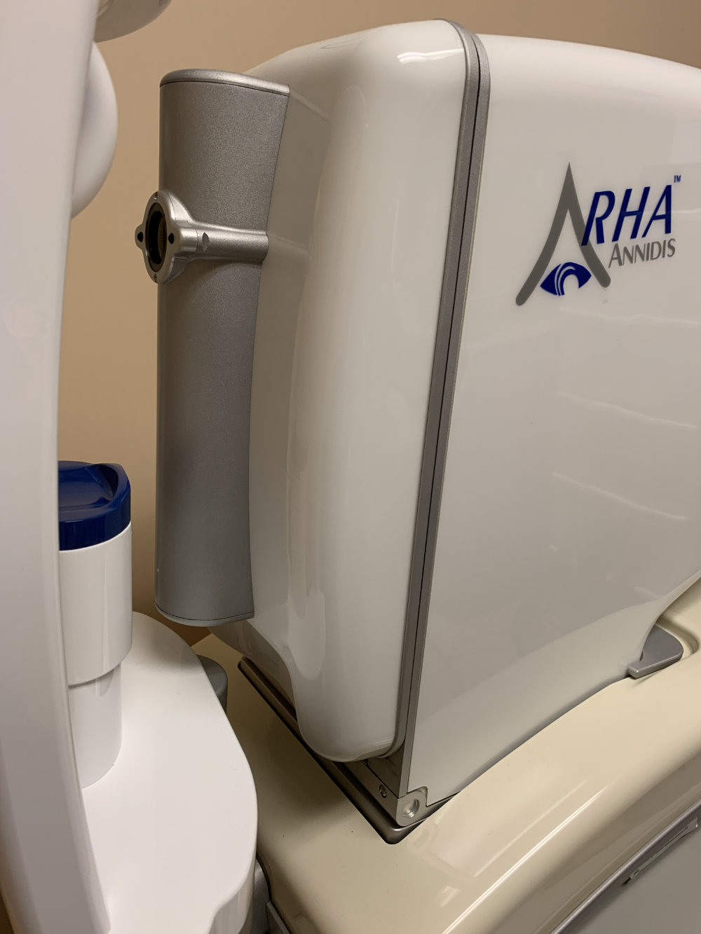
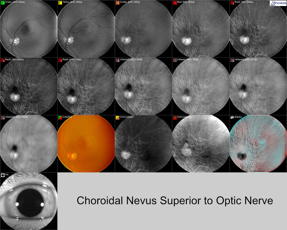
Multispectral retinal imaging is a relatively new technology that allows our eye doctor to see the different layers of the retina. This can help us diagnose and monitor different retinal diseases.
Retinal diseases can lead to vision loss, so it's important to diagnose and treat them as early as possible. Multispectral retinal imaging can also be used to monitor the progression of a retinal disease and track the effectiveness of treatment.
Multispectral retinal imaging is performed using a special camera that captures images of the retina at different wavelengths of light. These images can then be compared to each other to show pathology in different layers of the retina. This can help doctors to diagnose retinal diseases, such as macular degeneration, diabetic retinopathy, choroidal nevi and glaucoma.

• Individual narrow band multi spectral images can be optimized to show retinal features more clearly for each spectral wavelength band.
• The unique annular optical imaging system, combined with pupil centered illumination, results in reduced depth of field, enhancing both the visibility of the target structures, and the sensitivity to angular variations reducing reflections and light scattering.
• Multiple wavelengths combined with wavelength dependent choroidal absorption allows control of the degree of back-lighting from scleral reflection.
• Multiple closely spaced wavelengths in key spectral regions ensure images with maximum feature discernibility for the targeted structures across a wide range of patient pigmentation levels.
• Long wavelength bands probe through dense pigment to provide images of the deep retina and choroid.
• The longer wavelength bands experience less scattering when penetrating diffusive media such as cataracts and clouded vitreous.
• Annular imaging allows internal angular filtering for optical processing, such as creation of stereo pairs.
• Multiple wavelengths with differential penetration provide the ability to “see though” an overlapping structure to another beneath it.
• Trans-scleral choroidal illumination produces detailed, non-invasive, views of the choroid, comparable to those of ICG (indocyanine green) angiography.
• Multi-image computer processing can synthesize additional information, such as oxyhemoglobin/deoxyhemoglobin based contrast, not visible in the source images.
