Precorneal tear film after corneal collagen cross-linking in keratoconus

Purpose: To study the condition of precorneal tear film in keratoconus patients after corneal collagen cross-linking (CXL).

Material and methods: The study enrolled 24 patients (30 eyes) with stages I-III progressive keratoconus. All study patients underwent standard (Dresden) protocol CXL. Patients were examined before the surgery and 1 week, 1 month, and 3 months postoperatively. The condition of precorneal tear film was evaluated based on the results of Norn test (the tear film break-up time), Schirmer test, Jones test, optical coherence tomography based lacrimal meniscometry, and improved tearscopy.

Results: One month after CXL, patients with keratoconus experienced transient changes in the precorneal tear film condition – the tear film break-up time was reduced, the tear secretion was elevated, the lacrimal meniscus biometry parameters were increased, and the thickness of the tear film lipid layer was decreased.

Conclusion: Corneal changes resulting from CXL lead to transient disorders of the mucin and lipid layers of the precorneal tear film, which requires adequate tear substitute therapy in the postoperative period.

Keywords: cross-linking; dry eye; keratoconus; tear film.

Voronin GV, Bubnova IA, Averich VV, Sarkisova KG. Sostoyanie prekorneal’noi sleznoi plenki posle krosslinkinga rogovichnogo kollagena pri keratokonuse (predvaritel’noe soobshchenie) [Precorneal tear film after corneal collagen cross-linking in keratoconus (preliminary report)]. Vestn Oftalmol. 2021;137(5. Vyp. 2):224-230. Russian. doi: 10.17116/oftalma2021137052224. PMID: 34669331.

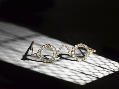
Sotheby’s Spectacle Auction

October 27, 2021 Live Auction in London. Arts of the Islamic World & India including Fine Rugs and Carpets.

A statement by Sotheby’s said that a diamond and an emerald were shaped into the two spectacles.

“The quality and purity of the gemstones is extraordinary and stones of this size would no doubt have been the reserve of an emperor,” it added.

The diamond lenses – cleaved as a pair from a single natural diamond – are thought to be from the mines of Golconda in southern India. The teardrop-shaped emeralds originate from a single natural Colombian emerald.

“While ordinary lenses merely function to improve sight, these filters were aids for spiritual enlightenment – with diamonds thought to illuminate and emeralds believed to have held miraculous powers to heal and to ward off evil,” Sotheby’s said.

According to the auction house, the “most famous evocation” of such glasses in history and mythology can be found in Pliny the Elder’s “Natural History”, which recounts the ancient Roman Emperor Nero observing gladiatorial contests through the surface of a precious green stone.

Nero’s tutor, Seneca, was an expert in light refraction, mirrors, and optics, and those were thought to be one of the first-ever spectacles, the statement said.

https://www.bbc.com/news/world-asia-india-58825741

Lot 213. A pair of Mughal spectacles set with emerald lenses, in diamond-mounted frames, India, lenses circa 17th century, frames 19th century.

View full screen - View 1 of Lot 213. A pair of Mughal spectacles set with emerald lenses, in diamond-mounted frames, India, lenses circa 17th century, frames 19th century.

Lot 214. A pair of Mughal spectacles set with diamond lenses, in diamond-mounted frames, India, lenses circa 17th century, frames 19th century.

View full screen - View 1 of Lot 214. A pair of Mughal spectacles set with diamond lenses, in diamond-mounted frames, India, lenses circa 17th century, frames 19th century.

Estimate: 1,500,000 – 2,500,000 GBP each. $2,000,000 – $3,400,000 each.

NIH-funded study shows screening device accurately detects amblyopia (lazy eye)

A handheld screening device that detects subtle misalignment of the eyes accurately identifies children with amblyopia (lazy eye), according to a study published in the Journal of the American Association for Pediatric Ophthalmology and Strabismus.

“The findings suggest that pediatricians and other primary care providers could use the device to catch  amblyopia at an early age when it’s easier to treat,” said Michael F. Chiang, M.D., director of the National Eye Institute (NEI), which supported research and development of the device. NEI is part of the National Institutes of Health.

Amblyopia is impaired vision in one eye and it is the leading cause of preventable monocular (single eye) vision loss, affecting three of every 100 children in the U.S.

During early childhood, our developing brains learn how to take images from each eye and fuse them into a single image to produce vision. Amblyopia develops when misalignment of the eyes (strabismus) or decreased acuity in one eye interferes with the brain’s ability to process visual information from both eyes, causing it to favor one eye. Once a child is visually mature, vision lost in the weaker eye cannot be corrected with glasses or contact lenses.

Children with amblyopia can suffer from poor school performance and impairments in depth perception and fine motor skills such as handwriting and other hand-eye coordinated activities.

Treatment of amblyopia generally involves placing a patch over the good eye to improve vision in the weaker eye. Patching is less successful as children get older, making early detection crucial. However, this depends on timely diagnosis by the child’s doctor, and most pediatricians are equipped only for basic eye chart vision screening tests, which are not useful for detecting amblyopia in very young children.

The screening device works by assessing the eyes’ ability to fixate together. Held 14 inches from the eyes, the child fixates on a smiley face while the device simultaneously scans both retinas. The scan involves a polarized laser that probes nerve fibers in an area of the light-sensing retina called the fovea, which is important for central vision. Even a subtle misalignment of the foveas — called small-angle strabismus — can interfere with the brain’s ability to integrate images from both eyes. The device calculates a binocularity score that indicates whether the child requires referral to an eye health physician for further investigation.

For the study, 300 children, ages 2 to 6 years, with no known eye disorders were recruited during previously scheduled visits to two Kaiser Permanente Southern California pediatric clinics.

Two non-ophthalmic research associates who were trained how to use the device screened each child, and the results were compared against those from an eye examination performed by a pediatric ophthalmologist who was unaware of the device’s results.  The device detected all six cases (100% sensitivity) of amblyopia and/or strabismus that had been confirmed by the professional eye examination. The device also flagged an additional 45 children as possibly having amblyopia and/or strabismus who were later determined by the eye examination to be normal (85% specificity).

“In the hands of a trained user, it takes 28 seconds to sit the child down, perform the test, and record the results – provided that the child cooperates — which is an important consideration for its adoption in busy pediatric care settings,” said the study’s co-lead investigator, Shaival S. Shah, M.D., a pediatric ophthalmologist at Kaiser Permanente based in Tustin Ranch, California. The test itself requires just 2.5 seconds of a child’s attention.

A key limitation of the study was that the device was not compared with currently available photo-screening devices. Such devices detect differences in light reflexes in each eye and produce images that can help detect risk factors for amblyopia, but not amblyopia itself, which can result in both over and under detection of amblyopia.

By contrast, the pediatric vision scanner detects the presence of amblyopia as opposed to the risk factors of amblyopia, which may lead to fewer unnecessary referrals to pediatric ophthalmologists.

The pediatric vision screener tested in the study is a prototype of blinq. ®, a device that was cleared by the U.S. Food and Drug Administration in 2016.

The clinical trial identifier is NCT02536963. The study was supported by NEI grant R44EY025926 to Rebiscan, Inc. (doing business as Rebion), the Boston-based firm that markets blinq.®. Support was provided under the Small Business Innovation Research (SBIR) program, a funding mechanism that helps independently owned companies with 500 or fewer employees bring biomedical technologies to the market.

Co-founder of Rebion, David Hunter, M.D., Ph.D., ophthalmologist-in-chief at Boston Children’s Hospital, co-invented blinq.® with support from NEI grant R01EY012883.

For more information, visit the amblyopia page on the NEI website. To learn more about the screening device, see Vision screening device improves detection of “lazy eye”.

NEI leads the federal government’s research on the visual system and eye diseases. NEI supports basic and clinical science programs to develop sight-saving treatments and address special needs of people with vision loss. For more information, visit https://www.nei.nih.gov.   

About the National Institutes of Health (NIH): NIH, the nation’s medical research agency, includes 27 Institutes and Centers and is a component of the U.S. Department of Health and Human Services. NIH is the primary federal agency conducting and supporting basic, clinical, and translational medical research, and is investigating the causes, treatments, and cures for both common and rare diseases. For more information about NIH and its programs, visit www.nih.gov.

Vision for the Future Project: Screening impact on the prevention and treatment of visual impairments in public school children in São Paulo City, Brazil

Abstract

Objectives: Uncorrected refractive errors are the leading cause of visual impairment in children. In this cross-sectional retrospective study, we analyzed a social visual screening program for school children in São Paulo, Brazil, evaluated its impact on the prevention and treatment of children’s visual disabilities, and assessed its epidemiological outcomes to outline suggestions for its improvement.

Methods: First-grade children from public schools were submitted to prior visual screening by their teachers. Selected children were forwarded to the hospital’s campaigns for a second screening by ophthalmologists and treatment if needed. Data were analyzed for age, sex, visual acuity, biomicroscopy, refractive errors, ocular movement disorders, amblyopia, number of donated spectacles, and number of children forwarded to specialized care.

Results: A total of 1080 children were included with mean age of 6.24±0.45 years. Children with normal ophthalmological exam, 591 (54.7%; 95% confidence interval [CI]: 51.7%-57.7%) were dismissed and considered false-positives. Myopia, hyperopia, and astigmatism components were found in 164 (15.2%; CI: 13.1%-17.4%), 190 (17.6%; CI: 15.3%-20.0%), and 330 (30.5%; CI: 27.8%-33.4%) children, respectively. Amblyopia was diagnosed in 54 (5%; CI: 3.5%-6.4%) children, and 117 (10.8%; CI: 9.8%-12.8%) presented ocular movement disorders. A total of 420 glasses were donated.

Conclusion: Epidemiological findings for amblyopia and refractive errors are consistent with those of similar studies. The expressive number of diagnoses performed and number of glasses donated to underprivileged children depict the importance of such projects. New guidelines to improve their cost-effectiveness, such as professional training and community sensitization, are imperative.

Costa DRD, Debert I, Susanna FN, Falabreti JG, Polati M, Susanna Júnior R. Vision for the Future Project: Screening impact on the prevention and treatment of visual impairments in public school children in São Paulo City, Brazil. Clinics (Sao Paulo). 2021 Oct 1;76:e3062. doi: 10.6061/clinics/2021/e3062. PMID: 34614115.

Comparison between estimated and measured myopia progression in Hong Kong children without myopia control intervention

Abstract

Purpose: To compare myopia progression estimated by the Brien Holden Vision Institute (BHVI) Myopia Calculator with cycloplegic measures in Hong Kong children wearing single-vision distance spectacles over a 1- and 2-year period.

Methods: Baseline age, spherical equivalent refraction (SER) and ethnicity of control participants from previous longitudinal myopia studies were input into the BHVI Myopia Calculator to generate an estimate of the SER at 1 and 2 years. Differences between the measured and estimated SER (116 and 100 participants with 1- and 2-year subjective refraction data, respectively, and 111 and 95 participants with 1- and 2-year objective refraction, respectively) were analysed, and the measured SER compared with the 95% confidence interval (CI) of the estimated SER.

Results: In children aged 7-13 years, 36% progressed within the 95% CI of the Myopia Calculator’s estimate, whereas 33% became less myopic than predicted (range 0.31 to 1.92 D less at 2 years) and 31% became more myopic than predicted (range 0.25 to 2.33 D more myopic at 2 years). The average difference between the estimated and measured subjective or objective SER at 1 and 2 years of follow-up was not clinically significant (<0.25 D).

Conclusions: On average, the BHVI Myopia Calculator estimated SER was in close agreement with measured cycloplegic SER after 1 and 2 years of follow-up (mean differences < 0.25 D). However, the measured myopia progression only fell within the 95% CI of the estimated SER for 32%-38% of children, suggesting that the BHVI ‘without management’ progression data should be interpreted with caution. The inclusion of additional data, modified to include axial elongation, from longitudinal studies of longer duration with larger sample sizes and a range of racial backgrounds may improve the Calculator’s ability to predict future myopia progression for individual children.

Yang Y, Cheung SW, Cho P, Vincent SJ. Comparison between estimated and measured myopia progression in Hong Kong children without myopia control intervention. Ophthalmic Physiol Opt. 2021 Oct 1. doi: 10.1111/opo.12895. Epub ahead of print. PMID: 34596263.

https://pubmed.ncbi.nlm.nih.gov/34596263/